Bassett Collection of Stereoscopic Images of Human Anatomy
Dissection of anterior and lateral regions of neck
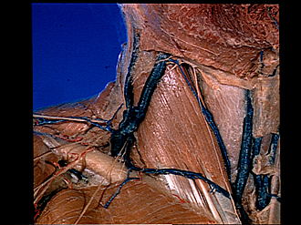
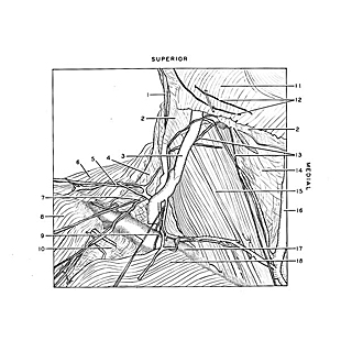
Right posterior cervical triangle; external jugular vein; supraclavicular nerves, anterolateral view
Image #72-7
KEYWORDS: Muscles and tendons, Peripheral nervous system, Vasculature.
Creative Commons
Stanford holds the copyright to the David L. Bassett anatomical images and has assigned Creative Commons license Attribution-Share Alike 4.0 International to all of the images.
For additional information regarding use and permissions, please contact the Medical History Center.
Dissection of anterior and lateral regions of neck
Right posterior cervical triangle; external jugular vein; supraclavicular nerves, anterolateral view
The platysma (11) has been resected and the underlying cervical fascia removed from the right side of the neck. The shoulder is to the left of the view and the chin to the upper right side.
- Great auricular nerve
- Fascia colli (external layer)
- External jugular vein (enlargement below pointer caused by valve sinuses within vein)
- Posterior cervical triangle
- Posterior supraclavicular nerve
- Trapezius muscle
- Medial supraclavicular nerve
- Deltoid muscle
- Anterior supraclavicular nerves
- Deltoid-pectoral trigonum
- Platysma (cut across below mandible)
- Cutaneous nerve filaments
- Cutaneus colli muscle (lower pointer indicates a long terminal branch, present bilaterally, which turned sharply and descended along the sternocleidomastoid muscle to the skin over the sternum)
- Fascia colli (middle layer over infrahyoid muscles in anterior cervical triangle)
- Sternocleidomastoid muscle
- Anterior jugular vein
- Clavicula
- Clavicular part of pectoralis major muscle
- [Legend above restored translation from Latin]


