
Bassett Collection of Stereoscopic Images of Human Anatomy
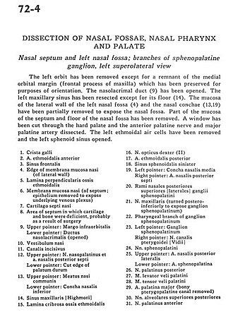
Dissection of nasal fossae, nasal pharynx, and palate
Nasal septum and left nasal fossa; branches of sphenopalatine ganglion, left superolateral view
Image #72-4
KEYWORDS: Bones cartilage joints, Face, Nose, Peripheral nervous system.
Creative Commons
Stanford holds the copyright to the David L. Bassett anatomical images and has assigned Creative Commons license Attribution-Share Alike 4.0 International to all of the images.

For additional information regarding use and permissions, please contact the Medical History Center.

Dissection of nasal fossae, nasal pharynx, and palate
Nasal septum and left nasal fossa; branches of sphenopalatine ganglion, left superolateral view
The left orbit has been removed except for the remnant of the medial orbital margin (frontal process of maxilla) which has been preserved for purposes of orientation. The nasolacrimal duct (9) has been opened. The left maxillary sinus has been resected except for its floor (14). The mucosa of the lateral wall of the left nasal fossa (4) and the nasal conchae (13,19) have been partially removed to expose the nasal fossa. Part of the mucosa of the septum and floor of the nasal fossa has been removed. A window has been cut through the hard palate and the anterior palatine nerve and major palatine artery dissected. The left ethmoidal air cells have been removed and the left sphenoid sinus opened.
- Crista galli
- Anterior ethmoidal artery
- Frontal sinus
- Edge of nasal mucosal membrane (of lateral wall)
- Perpendicular lamina of ethmoid bone
- Nasal mucosal membrane (of septum; epithelium removed to expose underlying venous plexus)
- Nasal septal cartilage
- Area of septum in which cartilage and bone were deficient, probably as a result of surgery
- Upper pointer: Infraorbital margo Lower pointer: Nasolacrimal duct (opened)
- Nasal vestibule
- Incisive canal
- Upper pointer: Nasopalatine nerve and posterior septal nasal artery Lower pointer: Cut edge of palatum durum
- Upper pointer: Communicating nasal meatus Lower pointer: Inferior nasal concha
- Maxillary sinus [Highmore]
- Cribrosal lamina of ethmoid bone
- Right optical nerve (II)
- Posterior ethmoid artery
- Left sphenoidal sinus
- Left pointer: Medial nasal concha Right pointer: Posterior nasal septal artery
- Rami nasales posteriores superiores [laterales] ganglii sphenopalatini
- Maxillary nerve (turned postero-inferiorly to expose sphenopalatine ganglion)
- Pharyngeal branch of sphenopalatine ganglion
- Left pointer: Sphenopalatine ganglion Right pointer: Pterygopalatine canal nerve [Vidius]
- Sphenopalatine nerves
- Upper pointer: Posterior lateral nasal artery Lower pointer: Sphenopalatine artery
- Posterior palatine nerve
- Palatine levator veli muscle
- Palatine tensor veli muscle
- Major palatine artery (bony pterygopalatine canal removed)
- Posterior superior alveolar nerves
- Anterior palatine nerve
- [Legend above restored translation from Latin]