Bassett Collection of Stereoscopic Images of Human Anatomy
Dissection of nose
Superficial nerves and blood vessels; transverse nasal muscle, left anterolateral view
Image #70-7
KEYWORDS: Connective tissue, Face, Muscles and tendons, Nose, Peripheral nervous system, Vasculature.
Creative Commons
Stanford holds the copyright to the David L. Bassett anatomical images and has assigned Creative Commons license Attribution-Share Alike 4.0 International to all of the images.
For additional information regarding use and permissions, please contact the Medical History Center.
Dissection of nose
Superficial nerves and blood vessels; transverse nasal muscle, left anterolateral view
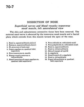
The skin and subcutaneous connective tissue have been removed. The external nasal nerve is obscured by the transverse nasal muscle and a fascial plane which extends from this muscle toward the apex of the nose.
- Branches of supratrochlear nerve left
- Branch supratrochlear nerve right
- Procerus muscle and base of nose
- Angular head of levator labii superioris muscle
- Transverse part of nasalis muscle
- External nasal branch infraorbital nerve
- Nasal insertion of angular head of levator labii superioris muscle
- Orbital part orbicularis oculi muscle
- Palpebral part of orbicularis oculi muscle
- Branches of supratrochlear nerve
- Branches of infratrochlear nerve
- Skin of margin of eyelid
- Angular artery
- Nasal branch of angular artery
- Branches of infraorbital nerve
- Infraorbital head of levator labii superioris muscle


