
Bassett Collection of Stereoscopic Images of Human Anatomy
Exploration of the brain from its basal aspect
Longitudinal fibers within septum pellucidum
Image #7-6
KEYWORDS: Brain, Telencephalon, Ventricules.
Creative Commons
Stanford holds the copyright to the David L. Bassett anatomical images and has assigned Creative Commons license Attribution-Share Alike 4.0 International to all of the images.

For additional information regarding use and permissions, please contact the Medical History Center.

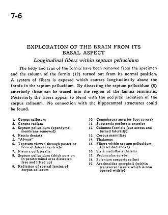
Exploration of the brain from its basal aspect
Longitudinal fibers within septum pellucidum
The body and crus of the fornix have been removed from the specimen and the column of the fornix (12) turned out from its normal position. A system of fibers is exposed which courses longitudinally above the fornix in the septum pellucidum. By dissecting the septum pellucidum (8) anteriorly these can be traced into the region of the lamina terminalis. Posteriorly the fibers appear to blend with the occipital radiation of the corpus callosum. No connection with the hippocampal structures could be found.
- Corpus callosum
- Corona radiata
- Septum pellucidum (ependymal membrane removed)
- Dentate fascia
- "Alveus"
- Tapetum viewed through posterior horn of lateral ventricle
- Collateral fissure
- Septum pellucidum (thick portion in paraterminal area dissected free and lifted up)
- Radiation of rostral lamina of corpus callosum
- Anterior commissure (cut across)
- Anterior perforated substance
- Fornix (column) (cut across and turned laterally)
- Mamillary body
- Thalamus
- Fibers within septum pellucidum (described above)
- Stria medullaris thalami
- Cerebral peduncle
- Corpus callosum (splenium)
- Arachnoid (within transverse fissure which is now opened widely)