Bassett Collection of Stereoscopic Images of Human Anatomy
Dissection of oral region
Relation of sublingual gland to tongue, left lateral view
Image #69-6
KEYWORDS: Cheek, Exocrine and endocrine, Face, Mouth, Muscles and tendons, Peripheral nervous system.
Creative Commons
Stanford holds the copyright to the David L. Bassett anatomical images and has assigned Creative Commons license Attribution-Share Alike 4.0 International to all of the images.

For additional information regarding use and permissions, please contact the Medical History Center.
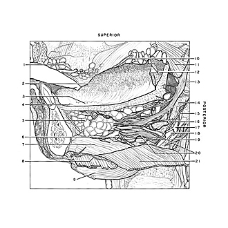
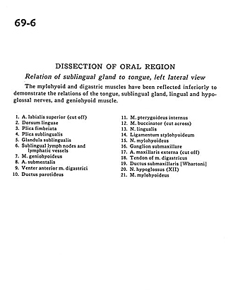
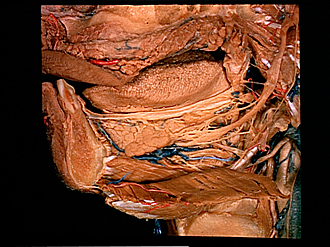
Dissection of oral region
Relation of sublingual gland to tongue, left lateral view
The mylohyoid and digastric muscles have been reflected inferiorly to demonstrate the relations of the tongue, sublingual gland, lingual and hypoglossal nerves, and geniohyoid muscle.
- Superior labial artery (cut off)
- Dorsum linguae
- Fimbriated fold (of tongue)
- Sublingual fold
- Sublingual gland
- Sublingual lymph nodes and lymphatic vessels
- Geniohyoid muscle
- Submental artery
- Anterior belly digastric muscle
- Parotid duct
- Internal pterygoid muscle
- Buccinator muscle (cut across)
- Lingual nerve
- Stylohyoid ligament
- Mylohyoid nerve
- Submandibular ganglion
- External maxillary artery (cut off)
- Tendon of digastric muscle
- Submandibular duct
- Hypoglossal nerve (XII)
- Mylohyoid muscle