Bassett Collection of Stereoscopic Images of Human Anatomy
Dissection of oral region
Oral cavity opened; nerves to mylohyoid and digastric muscles, left lateral view
Image #69-5
KEYWORDS: Cheek, Face, Mouth, Muscles and tendons, Peripheral nervous system.
Creative Commons
Stanford holds the copyright to the David L. Bassett anatomical images and has assigned Creative Commons license Attribution-Share Alike 4.0 International to all of the images.

For additional information regarding use and permissions, please contact the Medical History Center.
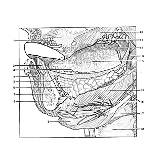
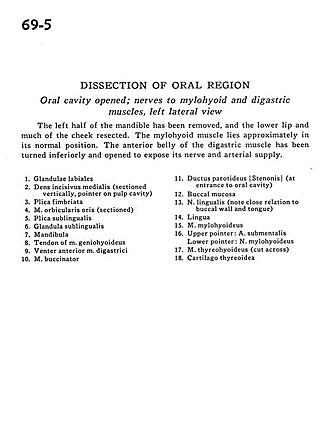
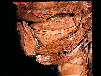
Dissection of oral region
Oral cavity opened; nerves to mylohyoid and digastric muscles, left lateral view
The left half of the mandible has been removed, and the lower lip and much of the cheek resected. The mylohyoid muscle lies approximately in its normal position. The anterior belly of the digastric muscle has been turned inferiorly and opened to expose its nerve and arterial supply.
- Labial glands
- Medial incisor (sectioned vertically, pointer on pulp cavity)
- Plica fimbriata
- Orbicularis oris muscle (sectioned)
- Sublingual fold
- Sublingual gland
- Mandible
- Tendon of geniohyoid muscle
- Anterior belly digastric muscle
- Buccinator muscle
- Parotid duct (at entrance to oral cavity)
- Buccal mucosa
- Lingual nerve (note close relation to buccal wall and tongue)
- Tongue
- Mylohyoid muscle
- Upper pointer: Submental artery Lower pointer: Mylohyoid nerve
- Thyrohyoid muscle (cut across)
- Thyroid cartilage