
Bassett Collection of Stereoscopic Images of Human Anatomy
Dissection of oral region
Lingual nerve; submaxillary ganglion and plexus, left lateral view
Image #69-4
KEYWORDS: Cheek, Connective tissue, Face, Mouth, Peripheral nervous system.
Creative Commons
Stanford holds the copyright to the David L. Bassett anatomical images and has assigned Creative Commons license Attribution-Share Alike 4.0 International to all of the images.

For additional information regarding use and permissions, please contact the Medical History Center.
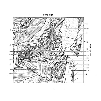
Dissection of oral region
Lingual nerve; submaxillary ganglion and plexus, left lateral view
The mylohyoid muscle has been retracted inferiorly and the lingual nerve freed of its connective tissue.
- Buccinator muscle and gingiva
- Sublingual gland
- Sublingual nerve (plexiform)
- Mandible (cut across)
- Submandibular gland (deep lobule)
- Tendon of digastric muscle
- Mylohyoid muscle (retracted inferiorly)
- Anterior belly digastric muscle
- Lingual nerve
- Mylohyoid nerve
- Branches of lingual nerve
- Upper pointer: Internal pterygoid muscle (pointer on area of insertion on angle of mandible) Lower pointer: Muscular branch of external maxillary artery
- External maxillary artery
- Submandibular ganglion
- Submandibular duct
- Stylohyoid muscle
- Submental artery
- Hypoglossal nerve (XII)
- External carotid artery
- Internal carotid artery
- Superior laryngeal nerve