Bassett Collection of Stereoscopic Images of Human Anatomy
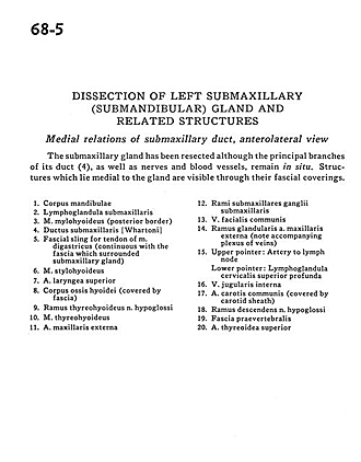
Dissection of left submaxillary (submandibular) gland and related structures
Medial relations of submaxillary duct, anterolateral view
Image #68-5
KEYWORDS: Exocrine and endocrine, Fascia and connective tissue, Peripheral nervous system, Throat, Vasculature.
Creative Commons
Stanford holds the copyright to the David L. Bassett anatomical images and has assigned Creative Commons license Attribution-Share Alike 4.0 International to all of the images.

For additional information regarding use and permissions, please contact the Medical History Center.
Dissection of left submaxillary (submandibular) gland and related structures
Medial relations of submaxillary duct, anterolateral view
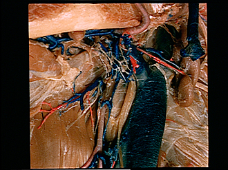
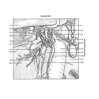
The submaxillary gland has been resected although the principal branches of its duct (4), as well as nerves and blood vessels, remain in situ. Structures which lie medial to the gland are visible through their fascial coverings.
- Body of mandible
- Submandibular lymph node
- Mylohyoid muscle (posterior border)
- Submandibular duct
- Fascial sling for tendon of digastric muscle (continuous with the fascia which surrounded submandibular gland)
- Stylohyoid muscle
- Superior laryngeal artery
- Body hyoid bone (covered by fascia)
- Thyrohyoid branch of hypoglossal nerve
- Thyrohyoid muscle
- External maxillary artery
- Submandibular branches of submandibular ganglion
- Common facial vein
- Glandular branch external maxillary artery (note accompanying plexus of veins)
- Upper pointer: Artery to lymph node Lower pointer: Superior deep cervical lymph node
- Internal jugular vein
- Common carotid artery (covered by carotid sheath)
- Descending branch hypoglossal nerve
- Prevertebral fascia
- Superior thyroid artery