Bassett Collection of Stereoscopic Images of Human Anatomy
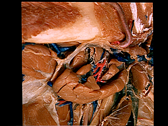
Dissection of left submaxillary (submandibular) gland and related structures
Intrinsic nerves and blood vessels, inferolateral view
Image #68-4
KEYWORDS: Exocrine and endocrine, Peripheral nervous system, Throat, Vasculature.
Creative Commons
Stanford holds the copyright to the David L. Bassett anatomical images and has assigned Creative Commons license Attribution-Share Alike 4.0 International to all of the images.

For additional information regarding use and permissions, please contact the Medical History Center.


Dissection of left submaxillary (submandibular) gland and related structures
Intrinsic nerves and blood vessels, inferolateral view
The lobules of the gland have been separated and some of them removed. The submaxillary duct has not been exposed.
- Platysma (cut off)
- Body of mandible and submandibular lymph node
- Fascia covering anterior belly digastric muscle
- Fascia which extends medial to submandibular gland (continuous with external layer of cervical fascia)
- Hyoid bone
- Thyrohyoid muscle
- Masseteric branch of external maxillary artery
- External maxillary artery
- Lymph vessel and submandibular lymph node
- Submental vein (cut off)
- Submandibular branches of submandibular ganglion
- Glandular branch external maxillary artery
- Submandibular gland
- Descending branch hypoglossal nerve
- Superior deep cervical lymph node
- Upper pointer: Carotid sheath covering external carotid artery Lower pointer: Superior thyroid artery