Bassett Collection of Stereoscopic Images of Human Anatomy
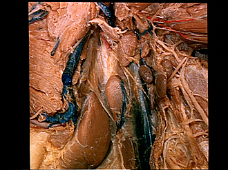
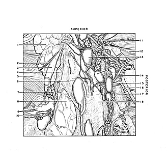
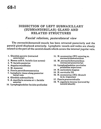
Dissection of left submaxillary (submandibular) gland and related structures
Facial relations, posterolateral view
Image #68-1
KEYWORDS: Exocrine and endocrine, Fascia and connective tissue, Muscles and tendons, Throat, Vasculature.
Creative Commons
Stanford holds the copyright to the David L. Bassett anatomical images and has assigned Creative Commons license Attribution-Share Alike 4.0 International to all of the images.

For additional information regarding use and permissions, please contact the Medical History Center.
Dissection of left submaxillary (submandibular) gland and related structures
Facial relations, posterolateral view
The sternocleidomastoid muscle has been retracted posteriorly and the parotid gland displaced anteriorly. Lymphatic vessels and nodes are closely related to the part of the carotid sheath which covers the internal jugular vein.
- Parotid gland (retracted anteriorly)
- Superficial branch facial nerve (cut across)
- Posterior facial vein
- Angle of mandible
- Masseter muscle
- Parotidomasseteric fascia
- Lymphatic tissue along posterior facial vein
- Submandibular gland
- External maxillary artery and anterior facial vein
- Deep facial lymph nodes
- Accessory nerve (XI) entering sternocleidomastoid muscle
- Sternocleidomastoid muscle (retracted posteriorly)
- Superior deep cervical lymph nodes
- Greater auricular nerve
- Cervical nerve III
- Accessory nerve (XI) (branch to trapezius muscle)
- Prevertebral fascia
- Internal jugular vein (covered by carotid sheath)