Bassett Collection of Stereoscopic Images of Human Anatomy
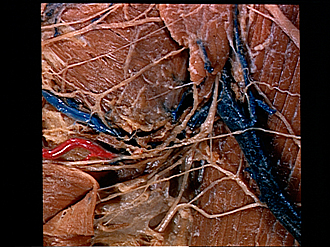
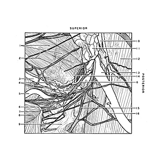
Dissection of left submaxillary (submandibular) gland and related structures
Branches of cervical plexus and facial nerve superficial to submaxillary gland
Image #67-7
KEYWORDS: Exocrine and endocrine, Fascia and connective tissue, Muscles and tendons, Peripheral nervous system, Throat.
Creative Commons
Stanford holds the copyright to the David L. Bassett anatomical images and has assigned Creative Commons license Attribution-Share Alike 4.0 International to all of the images.

For additional information regarding use and permissions, please contact the Medical History Center.
Dissection of left submaxillary (submandibular) gland and related structures
Branches of cervical plexus and facial nerve superficial to submaxillary gland
The left platysma (8) has been reflected anteriorly. The fascia which covered the sternocleidomastoid muscle (12) has been cut away but that which encases the submaxillary gland has been retained. Components of the extensive anastomosis between branches of the facial and cutaneous cervical nerves occupy much of the field.
- Masseter muscle and parotidomasseteric fascia
- Buccal branches of facial nerve
- Anterior facial vein
- Deep facial lymph nodes and lymph vessels
- External maxillary artery
- Marginal mandibular branches of facial nerve
- Submandibular lymph node
- Platysma (reflected)
- Superficial fascia (external layer)
- Remnant of superficial fascia which covered sternocleidomastoid muscle and continued superiorly as the parotidomasseteric fascia
- Parotid gland
- Sternocleidomastoid muscle
- External jugular vein
- Position of angle of mandible beneath masseter muscle
- Superficial branch facial nerve
- Branch cutaneous colli nerve (other branches visible superiorly)