
Bassett Collection of Stereoscopic Images of Human Anatomy
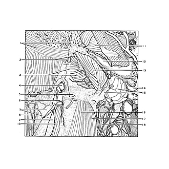
Dissection of submental region
Relations of digastric and mylohyoid muscles, submaxillary gland and hyoid bone, anteroinferior view
Image #67-6
KEYWORDS: Exocrine and endocrine, Muscles and tendons, Throat.
Creative Commons
Stanford holds the copyright to the David L. Bassett anatomical images and has assigned Creative Commons license Attribution-Share Alike 4.0 International to all of the images.

For additional information regarding use and permissions, please contact the Medical History Center.
Dissection of submental region
Relations of digastric and mylohyoid muscles, submaxillary gland and hyoid bone, anteroinferior view
The fascia has been cut away from the structures to the left of the midline.
- Mentalis muscle and transverse mental muscle
- Submental lymph nodes
- Upper pointer: Midline raphe Lower pointer: Mylohyoid muscle left
- Lymph node
- Body hyoid bone (covered by fibrous tissue)
- Anterior jugular vein
- Superficial fascia (external layer)
- Sternocleidomastoid muscle
- Superficial fascia (middle layer)
- Sternohyoid muscle right
- Platysma (reflected)
- Body of mandible
- Anterior belly digastric muscle
- External maxillary artery
- Submandibular gland (lower pointer indicates cut edge of investing fascia)
- Sternohyoid muscle left
- Omohyoid muscle
- Superior thyroid artery