Bassett Collection of Stereoscopic Images of Human Anatomy
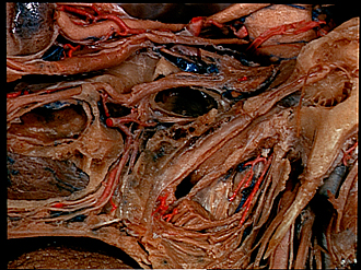
Dissection of pharynx from left lateral approach
Interior of auditory tube
Image #66-5
KEYWORDS: Ear, Pharynx, Throat.
Creative Commons
Stanford holds the copyright to the David L. Bassett anatomical images and has assigned Creative Commons license Attribution-Share Alike 4.0 International to all of the images.

For additional information regarding use and permissions, please contact the Medical History Center.


Dissection of pharynx from left lateral approach
Interior of auditory tube
The levator veli palatini muscle has been partially resected and the auditory tube opened.
- Optic nerve (II)
- Inferior rectus muscle
- Maxillary nerve (V)
- Sphenoid sinus
- Sphenopalatine ganglion
- Internal maxillary artery (cut across near termination)
- Maxillary sinus
- Mucosa of inferior nasal meatus
- Descending palatine artery
- Tendon of tensor veli palatini muscle (cut off)
- Bursa tensor veli palatini muscle
- Dorsum of tongue
- Superior pharyngeal constrictor muscle (upper pointer, pterygopharyngeus muscle; lower pointer, buccopharyngeus muscle)
- Upper pointer: Internal carotid artery Lower pointer: Oculomotor nerve (III)
- Tegmen tympani
- Chorda tympani
- Promontorium tympani and tympanic plexus
- Tensor tympani muscle
- Upper pointer: Bony part auditory tube (dark area on medial wall indicates position of carotid canal) Lower pointer: Isthmus auditory tube
- Lateral plate I cartilaginous auditory tube
- Scaphoid fossa sphenoid bone (site of origin of tensor veli palatini muscle)
- Upper pointer: Cartilaginous part auditory tube Lower pointer: Bony pharyngeal auditory tube
- Pharyngeal branch ascending pharyngeal artery
- Internal jugular vein
- Upper pointer: Alar fascia Lower pointer: Internal carotid artery
- Upper pointer: Pharyngobasilar fascia Lower pointer: Cut end of levator veli palatini muscle
- Styloid process
- Glossopharyngeal nerve (IX)
- Vagus nerve (X)