
Bassett Collection of Stereoscopic Images of Human Anatomy
Dissection of left infratemporal and pterygopalatine fossae
External pterygoid muscle, lateral view
Image #65-3
KEYWORDS: Muscles and tendons, Scalp, Vasculature.
Creative Commons
Stanford holds the copyright to the David L. Bassett anatomical images and has assigned Creative Commons license Attribution-Share Alike 4.0 International to all of the images.

For additional information regarding use and permissions, please contact the Medical History Center.

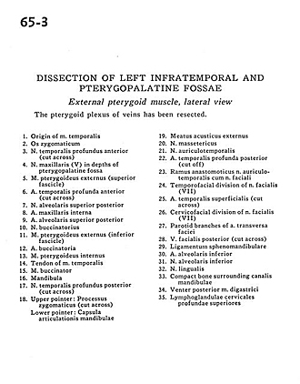
Dissection of left infratemporal and pterygopalatine fossae
External pterygoid muscle, lateral view
The pterygoid plexus of veins has been resected.
- Origin of temporalis muscle
- Zygomatic bone
- Anterior deep temporal nerve (cut across)
- Maxillary nerve (V) in depths of pterygopalatine fossa
- External pterygoid muscle (superior fascicle)
- Deep anterior temporal artery (cut across)
- Superior posterior alveolar nerve
- Internal maxillary artery
- Superior posterior alveolar artery
- Buccinator nerve
- External pterygoid muscle (inferior fascicle)
- Buccal artery
- Internal pterygoid muscle
- Tendon of temporalis muscle
- Buccinator muscle
- Mandible
- Posterior deep temporal nerve (cut across)
- Upper pointer: Zygomatic process (cut across) Lower pointer: Joint capsule of mandible
- External acoustic meatus
- Masseteric nerve
- Auriculotemporal nerve
- Deep posterior temporal artery (cut off)
- Anastomotic branch auriculotemporal nerve with facial nerve
- Temporofacial division of facial nerve (VII)
- Superficial temporal artery (cut across)
- Cervicofacial division of facial nerve (VII)
- Parotid branches of transverse facial artery
- Posterior facial vein (cut across)
- Sphenomandibular ligament
- Inferior alveolar artery
- Inferior alveolar nerve
- Lingual nerve
- Compact bone surrounding mandibular canal
- Posterior belly of digastric muscle
- Superior deep cervical lymph nodes