Bassett Collection of Stereoscopic Images of Human Anatomy
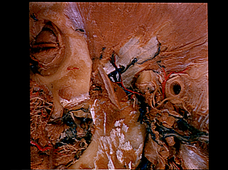
Dissection of left temporal region
Tendon of temporal muscle, lateral view
Image #64-5
KEYWORDS: Connective tissue, Muscles and tendons, Peripheral nervous system, Scalp.
Creative Commons
Stanford holds the copyright to the David L. Bassett anatomical images and has assigned Creative Commons license Attribution-Share Alike 4.0 International to all of the images.
For additional information regarding use and permissions, please contact the Medical History Center.
Dissection of left temporal region
Tendon of temporal muscle, lateral view
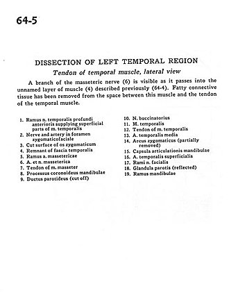
A branch of the masseteric nerve (6) is visible as it passes into the unnamed layer of muscle (4) described previously (64-4). Fatty connective tissue has been removed from the space between this muscle and the tendon of the temporal muscle.
- Branch anterior deep temporal nerve supplying superficial parts of temporalis muscle
- Nerve and artery in zygomaticofacial foramen
- Cut surface of zygomatic bone
- Remnant of temporal fascia
- Branch masseteric artery
- Masseteric nerve and artery
- Tendon of masseter muscle
- Coronoid process of mandible
- Parotid duct (cut off)
- Buccal nerve
- Temporalis muscle
- Tendon of temporalis muscle
- Middle temporal artery
- Zygomatic arch (partially removed)
- Joint capsule of mandible
- Superficial temporal artery
- Branches of facial nerve
- Parotid gland (reflected)
- Ramus of mandible


