Bassett Collection of Stereoscopic Images of Human Anatomy
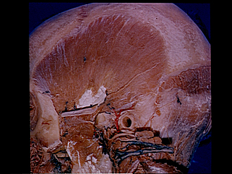
Dissection of left temporal region
Temporal muscle, lateral view
Image #64-4
KEYWORDS: Connective tissue, Muscles and tendons, Peripheral nervous system, Scalp.
Creative Commons
Stanford holds the copyright to the David L. Bassett anatomical images and has assigned Creative Commons license Attribution-Share Alike 4.0 International to all of the images.

For additional information regarding use and permissions, please contact the Medical History Center.


Dissection of left temporal region
Temporal muscle, lateral view
The zygomatic arch has been resected to reveal a separate layer of muscle (8) which arises from the inner surface of the arch and inserts on the coronoid process of the mandible. This muscle fascicle is supplied by a branch of masseteric nerve (12). A delicate sheet of muscle fibres related to the temporal fascia covers the main mass of the temporal muscle and is supplied by a branch (6) of the anterior deep temporal nerve.
- Coronal suture
- Frontal bone
- Temporal line
- Temporalis muscle
- Zygomaticotemporal branches of zygomatic nerve
- Nerve filament to superficial fascicle of temporalis muscle
- Remnant of temporal fascia which was attached to zygomatic arch
- Muscle origin from zygomatic arch (bone resected)
- Zygomaticofacial branch zygomatic nerve
- Malar surface zygomatic bone
- Position of coronoid process of mandible
- Masseteric nerve
- Tendon of masseter muscle
- Branches of facial nerve (reflected with parotid gland)
- Parietal bone
- Occipitalis muscle (cut across)
- Zygomatic process temporal bone (cut across)
- Origin of posterior auricular muscle
- Occipital branch of posterior auricular nerve
- Upper pointer: Middle temporal artery Lower pointer: Superficial temporal artery
- External acoustic meatus
- Auriculotemporal nerve (cut across)
- Lesser occipital nerve
- Parotid gland (reflected posteriorly)
- Parotid duct
- Sternocleidomastoid muscle and great auricular nerve