Bassett Collection of Stereoscopic Images of Human Anatomy
Dissection of left temporal region
Temporal fascia, lateral view
Image #64-2
KEYWORDS: Connective tissue, Scalp.
Creative Commons
Stanford holds the copyright to the David L. Bassett anatomical images and has assigned Creative Commons license Attribution-Share Alike 4.0 International to all of the images.

For additional information regarding use and permissions, please contact the Medical History Center.

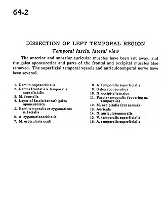
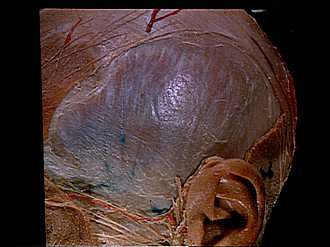
Dissection of left temporal region
Temporal fascia, lateral view
The anterior and superior auricular muscles have been cut away, and the galea aponeurotica and other parts of the frontal and occipital muscles also removed. The superficial temporal vessels and auriculotemporal nerve have been severed.
- Branches of supraorbital nerve
- Frontal branch of superficial temporal artery
- Frontalis muscle
- Layer of fascia beneath galea aponeurotica
- Temporal and zygomatic branches of facial nerve
- Zygomatico-orbital artery
- Orbicularis oculi muscle
- Superficial temporal artery
- Galea aponeurotica
- Greater occipital nerve
- Temporal fascia (covering temporalis muscle)
- Occipitalis muscle (cut across)
- Auricle
- Auriculotemporal nerve
- Superficial temporal vein
- Superficial temporal artery