Bassett Collection of Stereoscopic Images of Human Anatomy
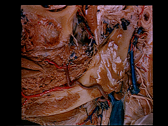
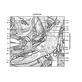
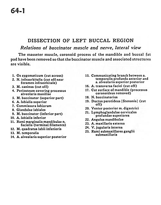
Dissection of left buccal region
Relations of buccinator muscle and nerve, lateral view
Image #64-1
KEYWORDS: Cheek, Face, Muscles and tendons, Peripheral nervous system.
Creative Commons
Stanford holds the copyright to the David L. Bassett anatomical images and has assigned Creative Commons license Attribution-Share Alike 4.0 International to all of the images.

For additional information regarding use and permissions, please contact the Medical History Center.
Dissection of left buccal region
Relations of buccinator muscle and nerve, lateral view
The masseter muscle, coronoid process of the mandible and buccal fat pad has been removed so that the buccinator muscle and associated structures are visible.
- Zygomatic bone (cut across)
- Infraorbital nerve (cut off near infraorbital foramen)
- Depressor anguli oris (cut off)
- Periosteum covering alveolar process of maxilla
- Buccinator muscle (superior part)
- Superior labial artery
- Labial commissure
- Labial glands
- Buccinator muscle (inferior part)
- Inferior labial artery
- Marginal mandibular branches facial nerve (terminal filaments)
- Depressor labii inferioris muscle
- Temporalis muscle
- Superior posterior alveolar artery
- Communicating branch between deep anterior temporal artery and superior posterior alveolar artery
- Transverse facial artery (cut off)
- Cut surface of mandible (coronoid process removed)
- Buccal nerve
- Parotid duct (cut off)
- Posterior belly of digastric muscle
- Superior deep cervical lymph nodes
- Angle of mandible
- External maxillary artery
- Internal jugular vein
- Submandibular branches of submandibular ganglion