Bassett Collection of Stereoscopic Images of Human Anatomy
Dissection of left ear from posterior aspect
Posterior surface of petrous part of temporal bone
Image #62-2
KEYWORDS: Bones cartilage joints, Ear.
Creative Commons
Stanford holds the copyright to the David L. Bassett anatomical images and has assigned Creative Commons license Attribution-Share Alike 4.0 International to all of the images.

For additional information regarding use and permissions, please contact the Medical History Center.
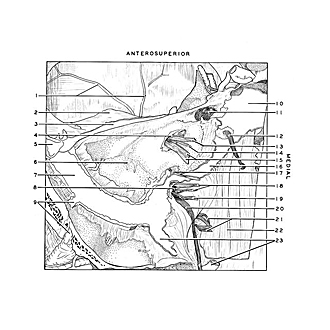
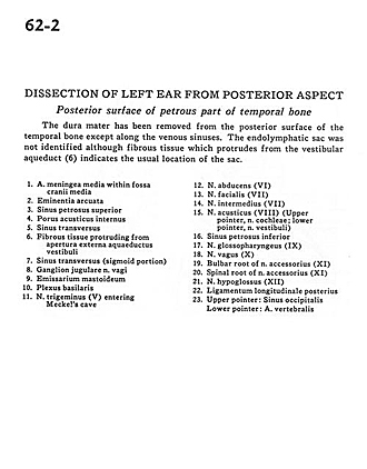
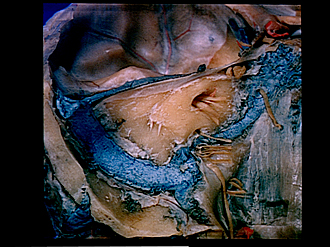
Dissection of left ear from posterior aspect
Posterior surface of petrous part of temporal bone
The dura mater has been removed from the posterior surface of the temporal bone except along the venous sinuses. The endolymphatic sac was not identified although fibrous tissue which protrudes from the vestibular aqueduct (6) indicates the usual location of the sac.
- Middle meningeal artery within middle cranial fossa
- Arcuate eminence
- Superior petrosal sinus
- Internal acoustic meatus
- Transverse sinus
- Fibrous tissue protruding from external aperture of vestibular aqueduct
- Transverse sinus (sigmoid portion)
- Jugular ganglion of vagus nerve
- Mastoid emissary
- Basilar plexus
- Trigeminal nerve (V) entering Meckel's cave
- Abducens nerve (VI)
- Facial nerve (VII)
- Nervous intermedius of (VII)
- Vestibulocochlear nerve (VIII) (upper pointer, cochlear part; lower pointer, vestibular part)
- Inferior petrosal sinus
- Glossopharyngeal nerve (IX)
- Vagus nerve (X)
- Bulbar root of accessory nerve (XI)
- Spinal root of accessory nerve (XI)
- Hypoglossal nerve (XII )
- Posterior longitudinal ligament
- Upper pointer: Occipital sinus Lower pointer: Vertebral artery