Bassett Collection of Stereoscopic Images of Human Anatomy
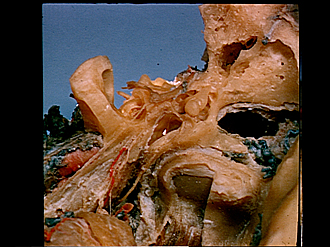
Dissection of left ear from superior aspect
Tympanic membrane
Image #61-7
KEYWORDS: Ear.
Creative Commons
Stanford holds the copyright to the David L. Bassett anatomical images and has assigned Creative Commons license Attribution-Share Alike 4.0 International to all of the images.

For additional information regarding use and permissions, please contact the Medical History Center.


Dissection of left ear from superior aspect
Tympanic membrane
- Facial nerve (VII)
- Capitulum of stapes
- Vestibule (opened)
- Superior pyramidal angle
- Vestibulocochlear nerve (vestibular part)
- Vestibulocochlear nerve (cochlear part)
- Osseous spiral lamina
- Modiolus: Upper pointer indicates scala vestibuli of middle turn; lower pointer indicates scala tympani of apical turn)
- Upper pointer: Tendon of tensor tympani muscle Lower pointer: Manubrium of malleus
- Major superficial petrosal nerve
- Minor superficial petrosal nerve
- Internal carotid artery within carotid canal
- Outer surface of auditory tube (Eustachian)
- Middle meningeal artery and mandibular nerve
- Incus
- Upper pointer: Incus-malleolar articulation Lower pointer: Malleus
- Flaccid part of tympanic membrane
- Anterior malleolar fold
- Pars tensa tympanic membrane
- Upper pointer: Limbus (border) of tympanic membrane Lower pointer: Jugular walls of tympanum
- Chorda tympani
- Capitulum condyloid process of mandible