Bassett Collection of Stereoscopic Images of Human Anatomy
Dissection of left ear from superior aspect
Relation of ear to temporomandibular joint, anterosuperior view
Image #61-4
KEYWORDS: Bones cartilage joints, Ear.
Creative Commons
Stanford holds the copyright to the David L. Bassett anatomical images and has assigned Creative Commons license Attribution-Share Alike 4.0 International to all of the images.

For additional information regarding use and permissions, please contact the Medical History Center.
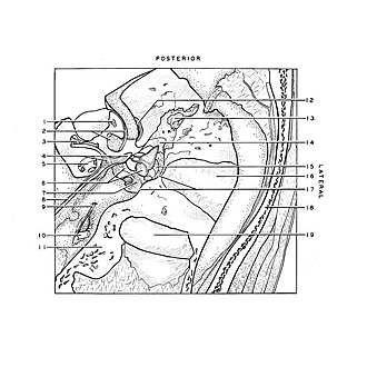
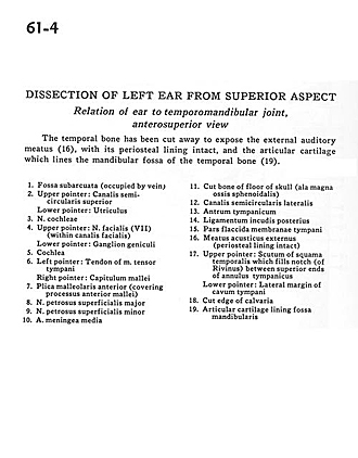
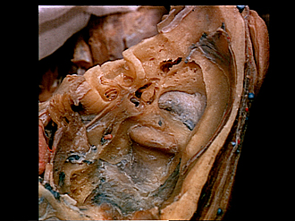
Dissection of left ear from superior aspect
Relation of ear to temporomandibular joint, anterosuperior view
The temporal bone has been cut away to expose the external auditory meatus (16), with its periosteal lining intact, and the articular cartilage which lines the mandibular fossa of the temporal bone (19).
- Subarcuate fossa (occupied by vein)
- Upper pointer: Superior semicircular canal Lower pointer: Utricle
- Vestibulocochlear nerve (cochlear part)
- Upper pointer: Facial nerve (VII) (within facial canal) Lower pointer: Geniculate ganglion
- Cochlea
- Left pointer: Tendon of tensor tympani muscle Right pointer: Capitulum of malleus
- Anterior malleolar fold (covering anterior process malleus)
- Greater superficial petrosal nerve
- Lessor superficial petrosal nerve
- Middle meningeal artery
- Cut bone of floor of skull (greater wing of sphenoid bone)
- Lateral semicircular canal
- Tympanic antrum
- Posterior ligament of incus
- Flaccid part of tympanic membrane
- External acoustic meatus (periosteal lining intact)
- Upper pointer: Scutum of temporal bone (squamous part) which fills notch (of Rivinus) between superior ends of tympanic annulus Lower pointer: Lateral margin of tympanic cavity
- Cut edge of calvaria
- Articular cartilage lining mandibular fossa