Bassett Collection of Stereoscopic Images of Human Anatomy
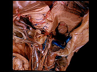
Contents of left jugular foramen
Superolateral view
Image #60-7
KEYWORDS: Bones cartilage joints, Ear, Peripheral nervous system.
Creative Commons
Stanford holds the copyright to the David L. Bassett anatomical images and has assigned Creative Commons license Attribution-Share Alike 4.0 International to all of the images.

For additional information regarding use and permissions, please contact the Medical History Center.

Contents of left jugular foramen
Superolateral view
The jugular foramen has been opened by cutting away its lateral wall. The internal jugular vein has been removed and its main tributaries, the sigmoid sinus (23) and the inferior petrosal sinus (12), cut off to expose the glossopharyngeal (13), vagus (24) and accessory (22) nerves which descend through the foramen. The dense fibrous tissue which surrounded these structures has been cleared away. The left cerebellar hemisphere has been partially removed.
- Upper pointer: Optic tract Lower pointer: Base of peduncle
- Internal carotid artery
- Posterior cerebral artery
- Clivus
- Upper pointer: Pons Lower pointer: Trigeminal nerve (V)
- Abducens nerve (VI)
- Sphenoid sinus
- Internal carotid plexus
- Upper pointer: Geniculate ganglion facial nerve (VII) Lower pointer: Vidian nerve (of pterygoid canal)
- Deep petrosal nerve
- Upper pointer: Cochlea (cut across) Lower pointer: Rectus capitis anterior muscle
- Upper pointer: Inferior petrosal sinus Lower pointer: Internal carotid nerve
- Glossopharyngeal nerve (IX)
- Superior cervical ganglion
- Cerebellum
- Superior cerebellar artery
- Dentate nucleus
- Cerebellar tonsil right
- Upper pointer: Brachium of pons (cut across) Lower pointer: Medulla oblongata
- Vestibulocochlear nerve (VIII)
- Roots of glossopharyngeal and vagus nerves
- Accessory nerve (XI)
- Transverse sinus (sigmoid part)
- Jugular ganglion of vagus nerve
- Medial wall of jugular foramen
- Mastoid cells
- Right pointer: Auricular branch vagus nerve (note communicating branch from glossopharyngeal nerve) Left pointer: Tympanic nerve
- Petrosal ganglion of glossopharyngeal nerve (superior ganglion absent in this specimen)
- Nodose ganglion of vagus nerve
- Hypoglossal nerve (XII)
- Rectus capitis lateralis muscle