Bassett Collection of Stereoscopic Images of Human Anatomy
Dissection of ear from lateral aspect
Medial wall of left tympanic cavity
Image #60-5
KEYWORDS: Bones cartilage joints, Ear.
Creative Commons
Stanford holds the copyright to the David L. Bassett anatomical images and has assigned Creative Commons license Attribution-Share Alike 4.0 International to all of the images.
For additional information regarding use and permissions, please contact the Medical History Center.
Dissection of ear from lateral aspect
Medial wall of left tympanic cavity
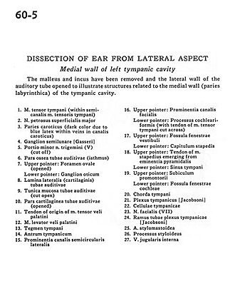
The malleus and incus have been removed and the lateral walls of the auditory tube opened to illustrate structures related to the medial wall (paries labyrinthica) of the tympanic cavity.
- Tensor tympani muscle (within semicircular canal)
- Major superficial petrosal nerve
- Carotid wall (dark color due to blue latex within veins in carotid canal)
- Semilunar ganglion
- Minor portion trigeminal nerve (V) (cut off)
- Bony part auditory tube (isthmus)
- Upper pointer: Foramen ovale (opened) Lower pointer: Otic ganglion
- Lateral plate (cartilaginous) auditory tube
- Mucosa of auditory tube (cut open)
- Cartilaginous part auditory tube (opened)
- Tendon of origin of Tensor veli palatini muscle
- Levator veli palatini muscle
- Tegmen tympani
- Tympanic antrum
- Promontory semicircular canal
- Upper pointer: Prominence of facial canal Lower pointer: Cochlearform process (with tendon of tensor tympani muscle cut across)
- Upper pointer: Fenestrated vestibular fossa Lower pointer: Capitulum of stapes
- Upper pointer: Tendon of stapedius muscle emerging from pyramidal eminence Lower pointer: Tympanic sinus
- Upper pointer: Promontory Lower pointer: Fenestrated cochlear fossa
- Chorda tympani
- Tympanic plexus
- Tympanic cells
- Facial nerve (VII)
- Tubal branch of tympanic plexus
- Stylomastoid artery
- Styloid process
- Internal jugular vein


