Bassett Collection of Stereoscopic Images of Human Anatomy
Dissection of ear from lateral aspect
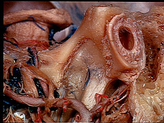
Petrotympanic fissure; relation of cartilaginous part of left external auditory meatus to tympanic part of temporal bone
Image #60-2
KEYWORDS: Bones cartilage joints, Brain, Diencephalon, Ear.
Creative Commons
Stanford holds the copyright to the David L. Bassett anatomical images and has assigned Creative Commons license Attribution-Share Alike 4.0 International to all of the images.

For additional information regarding use and permissions, please contact the Medical History Center.

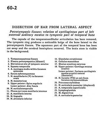
Dissection of ear from lateral aspect
Petrotympanic fissure; relation of cartilaginous part of left external auditory meatus to tympanic part of temporal bone
The capsule of the temporomandibular articulation has been removed. The tympanic ring produces a noticeable bulge of the bone lateral to the petrotympanic fissure. The squamous part of the temporal bone has been cut away and the cerebral hemisphere removed. The brain stem is visible in the background.
- Tympanosquamous fissure
- Petrotympanic fissure
- Elevation at site of tympanic annulus
- Temporal bone (squamous part)
- Middle meningeal artery within foramen spinosum
- Sphenosquamous suture
- Mandibular nerve (V) in foramen ovale
- Masseteric nerve
- Upper pointer: Angular spine Lower pointer: Alar fascia
- Auriculotemporal nerve
- Internal maxillary nerve plexus
- Internal maxillary artery
- Lingual nerve
- Inferior alveolar nerve
- Ceruminous gland
- Mastoid cells
- External acoustic meatus
- Upper pointer: Cartilaginous acoustic meatus Lower pointer: Incisure of external cartilaginous acoustic meatus
- Facial nerve (VII) at exit from stylomastoid foramen
- Styloid process
- Posterior auricular nerve (displaced)
- Superficial temporal artery
- Lymph node
- Digastric muscle
- Posterior auricular artery