Bassett Collection of Stereoscopic Images of Human Anatomy
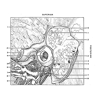
Dissection of ear from lateral aspect
Relation of left mastoid air cells to external auditory meatus
Image #60-1
KEYWORDS: Bones cartilage joints, Ear.
Creative Commons
Stanford holds the copyright to the David L. Bassett anatomical images and has assigned Creative Commons license Attribution-Share Alike 4.0 International to all of the images.
For additional information regarding use and permissions, please contact the Medical History Center.
Dissection of ear from lateral aspect
Relation of left mastoid air cells to external auditory meatus
The auricle has been cut away and the mastoid air cells opened. In this specimen pneumatization extended well above the petrosquamous fissure (12).
- Temporal fossa
- Upper pointer: Auricular cartilage (cut across) Lower pointer: Skin of external acoustic meatus
- Zygomatic process temporal bone (cut across)
- Upper pointer: External acoustic meatus Lower pointer: Cartilaginous acoustic meatus (covered by membrane)
- Upper pointer: Masseteric nerve Lower pointer: Articular disc of mandible
- Auriculotemporal nerve
- Upper pointer: Pterygoid venous plexus Lower pointer: Middle meningeal artery
- Superficial temporal artery
- Internal maxillary artery
- Middle temporal artery
- Dura mater exposed in dissected area of temporal bone
- Petrosquamous fissure
- Mastoid cells (opened)
- Splenius capitis muscle (cut away)
- Auricular branch vagus nerve (cut off)
- Posterior auricular nerve
- Upper pointer: Anastomotic branch auriculotemporal nerve with facial nerve Lower pointer: Facial nerve
- Upper pointer: Mastoid process Lower pointer: Tendon of sternocleidomastoid muscle


