Bassett Collection of Stereoscopic Images of Human Anatomy
Exploration of the brain from its basal aspect
Anterior limb of internal capsule, anterior commissure and parolfactory area
Image #6-3
KEYWORDS: Brain, Diencephalon, Telencephalon.
Creative Commons
Stanford holds the copyright to the David L. Bassett anatomical images and has assigned Creative Commons license Attribution-Share Alike 4.0 International to all of the images.

For additional information regarding use and permissions, please contact the Medical History Center.


Exploration of the brain from its basal aspect
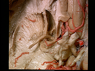
Anterior limb of internal capsule, anterior commissure and parolfactory area
The head of the caudate nucleus has been scraped away to reveal more clearly the fibers of the anterior limb of the internal capsule. These pass forward in discret bundles through the confluent parts of the caudate nucleus and putamen, the whole being named the "corpus striatum." Relations of the anterior commissure, anterior perforated substance and parolfactory area (now partly exposed by dissection) may also be seen.
- Radiation of rostral lamina of corpus callosum
- Space left by removal of head of caudate nucleus
- Frontal part internal capsule
- Parolfactory area (dissected)
- Subcallosal gyrus (dissected from within)
- Anterior commissure
- Anterior perforated substance
- Ansa lenticularis
- Cerebral peduncle
- Straight gyrus left
- Orbital branch of anterior cerebral artery
- Anterior cerebral artery
- Anterior communicating artery
- Olfactory tract
- Optic chiasm
- Internal carotid artery
- Infundibulum
- Interpeduncular fossa