
Bassett Collection of Stereoscopic Images of Human Anatomy
Dissection of ear from lateral aspect
Right auricular cartilage, lateral surface
Image #59-7
KEYWORDS: Bones cartilage joints, Ear.
Creative Commons
Stanford holds the copyright to the David L. Bassett anatomical images and has assigned Creative Commons license Attribution-Share Alike 4.0 International to all of the images.

For additional information regarding use and permissions, please contact the Medical History Center.
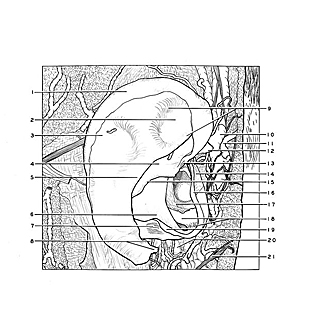
Dissection of ear from lateral aspect
Right auricular cartilage, lateral surface
The perichondrium has been removed.
- Helix
- Triangular fossa
- Scapha
- Cymba conchae
- Anthelix
- Cavum conchae
- Antitragicus muscle
- Upper pointer: Antitragicohelical fissure Lower pointer: Tail of helix
- Crura anthelicis
- Superficial temporal vein
- Superficial temporal artery
- Helical spine
- Helicis minor muscle
- Tragicus muscle
- Crus helicis
- Tragus
- Skin of external acoustic meatus
- Upper pointer: Cartilaginous acoustic meatus Lower pointer: Cartilaginous isthmus
- Antitragus
- Parotid gland
- Greater auricular nerve