
Bassett Collection of Stereoscopic Images of Human Anatomy
Dissection of ear from lateral aspect
Relation of right auricular cartilage to external auditory meatus; terminal branches of great auricular nerve
Image #59-5
KEYWORDS: Bones cartilage joints, Ear, Peripheral nervous system.
Creative Commons
Stanford holds the copyright to the David L. Bassett anatomical images and has assigned Creative Commons license Attribution-Share Alike 4.0 International to all of the images.

For additional information regarding use and permissions, please contact the Medical History Center.
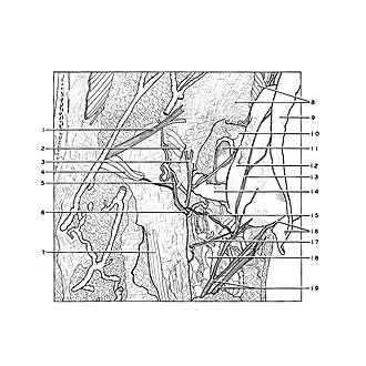
Dissection of ear from lateral aspect
Relation of right auricular cartilage to external auditory meatus; terminal branches of great auricular nerve
The posterior auricular muscle has been cut and the auricular cartilage retracted laterally.
- Lesser occipital nerve
- Posterior auricular muscle (cut across)
- Auricular branch vagus nerve
- Aberrant slip of posterior auricular muscle
- Occipital branch of posterior auricular nerve
- Posterior auricular nerve
- Sternocleidomastoid muscle (superficial fascia reflected)
- Upper pointer: Superior auricular muscle Lower pointer: Temporal fascia
- Helix
- Tragal plate
- External acoustic meatus (opened posteriorly)
- Conchal eminence
- Posterior branch greater auricular nerve
- Cartilaginous acoustic meatus
- Communication between posterior auricular nerve and greater auricular nerve
- Upper pointer: Antitragicohelical fissure Lower pointer: Tail of helix
- Branch facial nerve (to intrinsic muscles of auricle)
- Parotid gland
- Greater auricular nerve