
Bassett Collection of Stereoscopic Images of Human Anatomy
Microradiograph of eye; central optic pathways and related structures
Relations of optic pathways at base of brain.
Image #58A-6
KEYWORDS: Brain, Diencephalon, Exocrine and endocrine, Eye, Face, Midbrain, Peripheral nervous system.
Creative Commons
Stanford holds the copyright to the David L. Bassett anatomical images and has assigned Creative Commons license Attribution-Share Alike 4.0 International to all of the images.

For additional information regarding use and permissions, please contact the Medical History Center.
Microradiograph of eye; central optic pathways and related structures
Relations of optic pathways at base of brain.
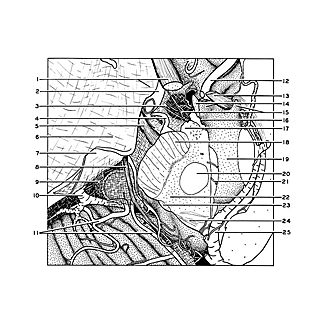
The vessels shown in the preceding view have been trimmed away and the left half of the optic chiasm has been removed to reveal the infundibulum of the hypophysis (15) extending downward through the opening in the diaphragm of the sella turcica (3). The recesses of the third ventricle that are closely related to the optic chiasm are indicated at 14 and 16. The tentorium has been cut and reflected to reveal the course of the trochlear nerve as it passes anteriorly around the mesencephalon.
- Upper pointer: Optic nerve (II) Lower pointer: Anterior clinoid process (covered by dura)
- Internal carotid artery
- Diaphragma sellae
- Posterior communicating artery
- Oculomotor nerve (III)
- Location of sinus cavernosus (covered by dura)
- Cerebral tentorium (reflected)
- Trochlear nerve (IV)
- Trigeminal nerve (V) (beneath arachnoid membrane)
- Superior cerebellar vein
- Branch of superior cerebellar artery
- Right anterior cerebral artery
- Anterior commissure
- Upper pointer: Optic chiasm (cut across in midline) Lower pointer: Optic recess
- Left pointer: Infundibulum Right pointer: Triangular recess
- Infundibular recess
- Mamillary body (cut across)
- Cerebral peduncle
- Third ventricle
- Nucleus ruber
- Arterial branch which entered brain stem through posterior perforated substance in interpeduncular fossa
- Position of medial lemniscus
- Brachium quadrigeminum inferius (cut across)
- Superior colliculus
- Left internal cerebral vein
- [Legend above restored translation from Latin]