Bassett Collection of Stereoscopic Images of Human Anatomy
Microradiograph of eye; central optic pathways and related structures
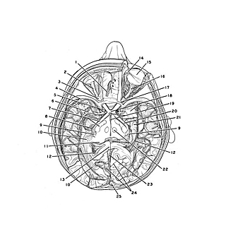
Optic pathways dissected in situ, viewed from above
Image #58A-2
KEYWORDS: Brain, Diencephalon, Eye, Face, Occipital lobe, Peripheral nervous system, Telencephalon.
Creative Commons
Stanford holds the copyright to the David L. Bassett anatomical images and has assigned Creative Commons license Attribution-Share Alike 4.0 International to all of the images.
For additional information regarding use and permissions, please contact the Medical History Center.
Microradiograph of eye; central optic pathways and related structures
Optic pathways dissected in situ, viewed from above
The visual pathways have been exposed bilaterally from the eyes to the calcarine cortex. The brain stem has been transected horizontally at the level of the optic tracts. The cerebral hemispheres have been dissected somewhat differently on the two sides. The optic radiation (10) has been preserved on the left side but has been divided on the right to expose the underlying inferior horn of the lateral ventricle. Within the right orbit the sheath of the optic nerve has been opened to expose the optic nerve. The sheath remains intact in the left orbit.
- Olfactory tract
- External vagina of optic nerve
- Anulus tendineus communis
- Vein cerebri media superficialis
- Upper pointer: Internal carotid artery Lower pointer: Anterior cerebral artery (cut off)
- Medial cerebral artery (within lateral cerebral sulcus)
- Upper pointer: Optical tract Lower pointer: Cerebral peduncle
- Lateral ventricular choroid plexus
- Lateral geniculate body (partially resected on right side)
- Optic radiation
- Pulvinar
- Lateral ventricle
- Splenium corporis callosi
- Oblique superior trochlear muscle
- Ocular bulb
- Optic nerve (exposed within sheath)
- Optic canal (partially opened to expose nerve)
- Upper pointer: Optic chiasm Lower pointer: Medial cranial fossa (uncus and medial part of temporal lobe removed)
- Tertiary ventricle (opened from supraoptic recess at upper pointer to communication with cerebral aqueduct posteriorly)
- Optic tract
- Hippocampus
- Nucleus ruber
- Inferior sagittal sinus (cut across)
- Calcarine sulcus (exposed by removal of cuneus on left; sectioned horizontally on right)
- Superior sagittal sinus
- [Legend above restored translation from Latin]


