Bassett Collection of Stereoscopic Images of Human Anatomy
Dissection of eye
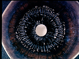
Ciliary body and iris, posterior view
Image #58-7
KEYWORDS: Eye, Face.
Creative Commons
Stanford holds the copyright to the David L. Bassett anatomical images and has assigned Creative Commons license Attribution-Share Alike 4.0 International to all of the images.
For additional information regarding use and permissions, please contact the Medical History Center.
Dissection of eye
Ciliary body and iris, posterior view
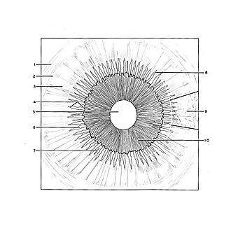
The lens has been removed to expose the iris and pupil. Remains of the vitreous body are visible at the periphery of the view (posterior to the ora serrata). The delicate hyloid membrane (8) is visible as a thin layer which covers the outer margin of the corona ciliaris but has been cut away medially. The cut edge is indicated by a white line in the drawing and the membrane is drawn only in a small area at the right (9).
- Optic (visual) part of retina
- Ora serrata
- Orbiculus ciliaris
- Corona ciliaris
- Pupil (posterior surface of cornea visible in background)
- Ciliary process
- Plica ciliaris
- Cut edge of hyaloid membrane
- Hyaloid membrane (drawn only in area within heavy lines)
- Posterior surface of iris


