Bassett Collection of Stereoscopic Images of Human Anatomy
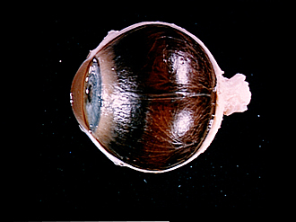
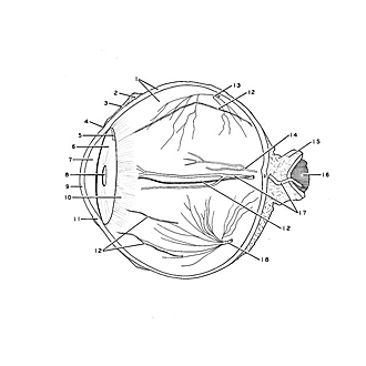
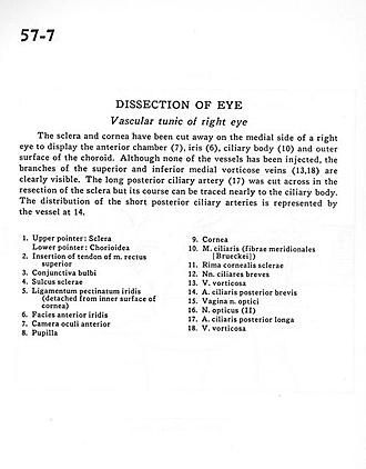
Dissection of eye
Vascular tunic of right eye
Image #57-7
KEYWORDS: Eye, Face, Peripheral nervous system, Vasculature.
Creative Commons
Stanford holds the copyright to the David L. Bassett anatomical images and has assigned Creative Commons license Attribution-Share Alike 4.0 International to all of the images.

For additional information regarding use and permissions, please contact the Medical History Center.
Dissection of eye
Vascular tunic of right eye
The sclera and cornea have been cut away on the medial side of a right eye to display the anterior chamber (7), iris(6), ciliary body(10) and outer surface of the choroid. Although none of the vessels has been injected, the branches of the superior and inferior medial vorticose veins (13,18) are clearly visible. The long posterior ciliary artery (17) was cut across in the resection of the sclera but its course can be traced nearly to the ciliary body. The distribution of the short posterior ciliary arteries is represented by the vessel at 14.
- Upper pointer: Sclera Lower pointer: Choroid
- Insertion of tendon of superior rectus muscle
- Bulbar conjunctiva
- Scleral sulcus
- Pectinate ligament of iridocorneal angle (detached from inner surface of cornea)
- Anterior surface of iris
- Anterior chamber of eye
- Pupil
- Cornea
- Ciliary muscle (meridional fibers)
- Corneo-scleral border (rim)
- Short diary nerves
- Vorticose vein
- Posterior ciliary artery (short)
- Sheath of optic nerve
- Optic nerve (II)
- Posterior ciliary artery (long)
- Vorticose vein