
Bassett Collection of Stereoscopic Images of Human Anatomy
Insertion of extraocular muscles
Left eye, anterior view
Image #57-5
KEYWORDS: Connective tissue, Eye, Face, Muscles and tendons.
Creative Commons
Stanford holds the copyright to the David L. Bassett anatomical images and has assigned Creative Commons license Attribution-Share Alike 4.0 International to all of the images.

For additional information regarding use and permissions, please contact the Medical History Center.
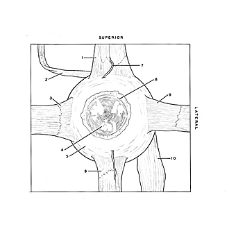

Insertion of extraocular muscles
Left eye, anterior view
The muscles have been elevated and stretched.
- Tendon of superior rectus muscle
- Tendon of superior oblique muscle (portion of tendon which originally passed through trochlea turned upward)
- Tendon of medial rectus muscle
- Cornea
- Sclera (conjunctiva and bulbar fascia removed)
- Tendon of Inferior rectus muscle
- Anterior ciliary artery
- Annulus conjunctivae overlying limbus corneae
- Tendon of lateral rectus muscle
- Inferior oblique muscle