Bassett Collection of Stereoscopic Images of Human Anatomy
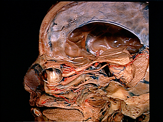
Dissection of left orbit from a lateral approach
General view of orbit, eye and optic pathway to lateral geniculate body
Image #56-5
KEYWORDS: Brain, Diencephalon, Eye, Face, Peripheral nervous system.
Creative Commons
Stanford holds the copyright to the David L. Bassett anatomical images and has assigned Creative Commons license Attribution-Share Alike 4.0 International to all of the images.

For additional information regarding use and permissions, please contact the Medical History Center.
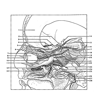

Dissection of left orbit from a lateral approach
General view of orbit, eye and optic pathway to lateral geniculate body
The eye has been sectioned in a sagittal plane and the sheath of the optic nerve cut away. The optic nerve (7) and the opthalmic artery(10) are visible passing through the optic canal which has been opened by removal of the anterior clinoid process. The sphenoid sinus extended into this process and has been cut open inferior to the opthalmic artery. The dissection of areas in the lower part of the view is shown in detail elsewhere.
- Falx cerebri
- Optic chiasm
- Sphenoidal border
- Olfactory tracts
- Upper pointer: Levator palpebrae superioris muscle Lower pointer: Superior rectus muscle
- Supraorbital margin (cut across)
- Upper pointer: Medial rectus muscle Lower pointer: Optic nerve (II)
- Upper pointer: Sclera (internal surface) Lower pointer: Cornea (somewhat distorted)
- Crystalline lens
- Ophthalmic artery
- Upper pointer: Inferior rectus muscle Lower pointer: Inferior oblique muscle (cut off)
- Infraorbital nerve
- Maxillary sinus (opened)
- Internal carotid artery right
- Third ventricle (opened)
- Inferior colliculus
- Lateral geniculate body
- Optic tract
- Oculomotor nerve (III)
- Internal carotid artery left
- Semilunar ganglion (trigeminal)
- Abducens nerve (VI)
- Major superficial petrosal nerve
- Sphenoid sinus (opened)
- Sphenopalatine ganglion
- Upper pointer: Auditory tube (Eustachian) (opened) Lower pointer: Levator veli palatini muscle
- Internal maxillary artery (pterygopalatine portion, cut off)
- Facial nerve emerging from stylomastoid foramen