Bassett Collection of Stereoscopic Images of Human Anatomy
Arteries of right orbital region of newborn
Lateromedial roentgenogram
Image #55-1
KEYWORDS: Eye, Face, Vasculature.
Creative Commons
Stanford holds the copyright to the David L. Bassett anatomical images and has assigned Creative Commons license Attribution-Share Alike 4.0 International to all of the images.
For additional information regarding use and permissions, please contact the Medical History Center.
Arteries of right orbital region of newborn
Lateromedial roentgenogram
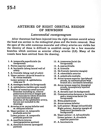
After thorotrast had been injected into the right common carotid artery the head was section in the midsagittal plane and the brain removed. Near the apex of the orbit numerous muscular and ciliary arteries are visible but the identity of these is difficult to establish except for a few muscular branches which continue as anterior ciliary arteries (5,9). Many of the vessels have been omitted from the drawing.
- Superficial temporal artery (in foreground)
- Superior wall orbit
- Lacrimal artery (along lateral wall of orbit)
- Frontal artery (along roof of orbit)
- Upper pointer: Arterial branch to medial rectus muscle Lower pointer: Ophthalmic artery (also see 7)
- Middle meningeal artery (in foreground)
- Ophthalmic artery (within optic canal)
- Region of numerous small arterial branches including posterior ciliary arteries (short) and long and muscular branches (these branches are derived from all major branches of the ophthalmic artery but are not included in the drawing)
- Arteries to inferior rectus muscle and inferior oblique muscle
- Upper pointer: Deep anterior temporal artery Lower pointer: Deep posterior temporal artery
- Internal maxillary artery (pterygopalatine portion)
- Internal maxillary artery (pterygoid portion)
- Transverse facial artery (in foreground)
- Supraorbital artery
- Frontal artery
- Frontal bone (anterior margin)
- Anterior ethmoidal artery
- Middle palpebral artery
- Superior tarsal arch (peripheral arcade, in communication with branch of lacrimal artery laterally)
- Superior tarsal arch (marginal arcade, incompletely injected laterally)
- Dorsal nasal artery (in background)
- Communication between lacrimal artery and transverse facial artery (in foreground)
- Palpebral rim
- Inferior tarsal arch
- Left pointer: Infraorbital margin Right pointer: Anterior margin of fossa of lacrimal sac (in background)
- Upper pointer: Infraorbital artery Lower pointer: Superior posterior alveolar artery
- Buccal artery


