Bassett Collection of Stereoscopic Images of Human Anatomy
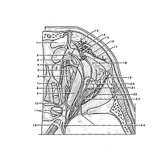
Dissection of right orbit from a superior approach
Relation of aponeurosis of levator palpebrae superioris muscle to tendon of superior oblique muscle and lacrimal gland
Image #54-2
KEYWORDS: Connective tissue, Exocrine and endocrine, Eye, Face, Muscles and tendons.
Creative Commons
Stanford holds the copyright to the David L. Bassett anatomical images and has assigned Creative Commons license Attribution-Share Alike 4.0 International to all of the images.
For additional information regarding use and permissions, please contact the Medical History Center.
Dissection of right orbit from a superior approach
Relation of aponeurosis of levator palpebrae superioris muscle to tendon of superior oblique muscle and lacrimal gland
The bone of the orbital margin has been resected and the fascia covering the levator palpebrae muscle removed. Subcutaneous structures of the supraorbital region (13-16) have been preserved along with the thickened rim of periorbita at the margin of the orbit (17). The superior orbital fissure (24) has been opened to display nerves which traverse it. The superior ophthalmic vein has been removed.
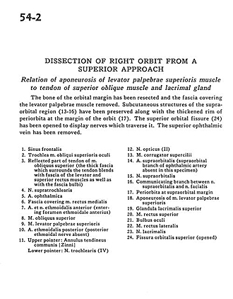
- Frontal sinus
- Superior oblique muscle
- Reflected part of tendon of superior oblique muscle (the thick fascia which surrounds the tendon blends with fascia of the levator and superior rectus muscles as well as with the bulbar fascia)
- Supratrochlear nerve
- Ophthalmic artery
- Fascia covering medial rectus muscle
- Anterior ethmoidal artery and nerve (entering anterior ethmoidal foramen)
- Superior oblique muscle
- Levator palpebrae superioris muscle
- Posterior ethmoidal artery (posterior ethmoidal nerve absent)
- Upper pointer: Common annular tendon Lower pointer: Trochlear nerve (IV)
- Optic nerve (II)
- Corrugator supercilii muscle
- Supraorbital artery (supraorbital branch of ophthalmic artery absent in this specimen)
- Supraorbital nerve
- Communicating branch between supraorbital nerve and facial nerve
- Periorbita at supraorbital margin
- Aponeurosis of levator palpebrae superioris muscle
- Superior lacrimal gland
- Superior rectus muscle
- Eyeball
- Lateral rectus muscle
- Lacrimal nerve
- Superior orbital fissure (opened)


