
Bassett Collection of Stereoscopic Images of Human Anatomy
Dissection of left orbit from an anterior approach
Inferior lacrimal part of orbicularis oculi muscle
Image #53-2
KEYWORDS: Connective tissue, Eye, Face, Muscles and tendons.
Creative Commons
Stanford holds the copyright to the David L. Bassett anatomical images and has assigned Creative Commons license Attribution-Share Alike 4.0 International to all of the images.

For additional information regarding use and permissions, please contact the Medical History Center.

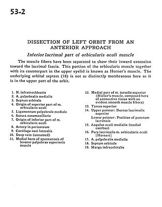
Dissection of left orbit from an anterior approach
Inferior lacrimal part of orbicularis oculi muscle
The muscle fibers have been separated to show their inward extension toward the lacrimal fascia. This portion of the orbicularis muscle together with its counterpart in the upper eyelid is known as Horner's muscle. The underlying orbital septum (18) is not as distinctly membranous here as it is in the upper part of the orbit.
- Infratrochlear nerve
- Middle palpebral artery
- Orbital septum
- Origin of superior part of orbicularis oculi muscle
- Medial palpebral ligament
- Nasomaxillary suture
- Origin of inferior part of orbicularis oculi muscle
- Artery in periosteum
- Lateral nasal cartilage
- Deep vein (unnamed)
- Medial horn of aponeurosis of levator palpebrae superioris muscle
- Medial part of superior tarsalis muscle (Müller's muscle, composed here of connective tissue with no evident smooth muscle fibers)
- Superior tarsus
- Upper pointer: Superior lacrimal duct Lower pointer: Position of lacrimal puncta
- Medial ocular angle (medial canthus)
- Lacrimal part orbicularis. oculi muscle
- Middle palpebral artery
- Orbital septum
- Infraorbital margin