
Bassett Collection of Stereoscopic Images of Human Anatomy
Dissection of left orbit from an anterior approach
Muscle fibers near medial angle of eyelids
Image #52-7
KEYWORDS: Connective tissue, Eye, Face.
Creative Commons
Stanford holds the copyright to the David L. Bassett anatomical images and has assigned Creative Commons license Attribution-Share Alike 4.0 International to all of the images.

For additional information regarding use and permissions, please contact the Medical History Center.
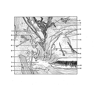

Dissection of left orbit from an anterior approach
Muscle fibers near medial angle of eyelids
Note that no fibers attach to the anterior surface of the medial palpebral ligament.
- Infratrochlear nerve
- Muscle fibers which joined frontalis muscle
- Periosteum covering nasal bone
- Area of origin of superior part of orbicularis oculi muscle
- Area of origin of inferior part of orbicularis oculi muscle
- Lacrimal part orbicularis oculi muscle
- Medial palpebral ligament
- Angular head of levator labii superioris muscle (cut across)
- Frontal nerve
- Corrugator supercilii muscle
- Supratrochlear nerve
- Orbital septum (note its downward extension deep to the orbicularis muscle toward an attachment on the posterior lacrimal crest)
- Aponeurosis of levator palpebrae superioris muscle (upper pointer, cutaneous insertion; lower pointer, tarsal insertion)
- Superior tarsus
- Middle palpebral artery
- Superior palpebral nerves
- Lacrimal duct
- Lacrimal lake
- Medial ocular angle