Bassett Collection of Stereoscopic Images of Human Anatomy
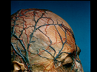
Scalp
Superficial nerves and blood vessels of scalp, anterolateral view
Image #51-6
KEYWORDS: Connective tissue, Muscles and tendons, Peripheral nervous system, Scalp, Vasculature.
Creative Commons
Stanford holds the copyright to the David L. Bassett anatomical images and has assigned Creative Commons license Attribution-Share Alike 4.0 International to all of the images.

For additional information regarding use and permissions, please contact the Medical History Center.

Scalp
Superficial nerves and blood vessels of scalp, anterolateral view
Arteries, veins and nerves which lie in the tela subcutanea have been exposed. Branches of the supraorbital nerve (1) lie mostly beneath the galea aponeurotica and consequently are exposed only near their terminations.
- Branches of supraorbital nerve
- Superficial temporal vein (posterior branch)
- Superficial temporal vein (anterior branch)
- Location of temporalis muscle beneath galea aponeurotica and temporal fascia
- Frontal branch superficial temporal artery
- Parietal branch superficial temporal artery
- Auriculotemporal nerve
- Superficial temporal artery
- Superficial fascia
- Galea aponeurotica
- Frontalis muscle
- Frontal vein
- Frontal artery (supraorbital artery faintly visible beneath frontalis muscle in a parallel, more lateral position)
- Supraorbital vein
- Anastomosis of superficial veins with middle temporal vein
- Angular vein
- Superior tarsus and orbicularis oculi muscle (cut away)