Bassett Collection of Stereoscopic Images of Human Anatomy
Floor of cranial cavity
Structures inferior to left middle cranial fossa (continued); maxillary and mandibular nerves
Image #51-5
KEYWORDS: Bones cartilage joints, Muscles and tendons, Peripheral nervous system, Vasculature.
Creative Commons
Stanford holds the copyright to the David L. Bassett anatomical images and has assigned Creative Commons license Attribution-Share Alike 4.0 International to all of the images.

For additional information regarding use and permissions, please contact the Medical History Center.
Floor of cranial cavity
Structures inferior to left middle cranial fossa (continued); maxillary and mandibular nerves
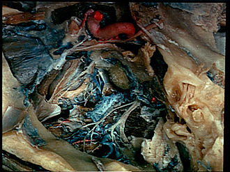
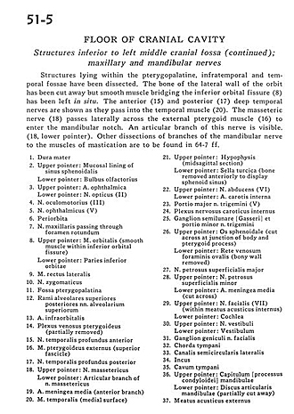
Structures lying within the pterygopalatine, infratemporal and temporal fossae have been dissected. The bone of the lateral wall of the orbit has been cut away but smooth muscle bridging the inferior orbital fissure (8) has been left in situ. The anterior (15) and posterior (17) deep temporal nerves are shown as they pass into the temporal muscle (20). The masseteric nerve (18) passes laterally across the external pterygoid muscle (16) to enter the mandibular notch. An articular branch of this nerve is visible. (18, lower pointer). Other dissections of branches of the mandibular nerve to the muscles of mastication are to be found in 64-7 ff.
- Dura mater
- Upper pointer: Mucosal lining of sphenoid sinus Lower pointer: Olfactory bulb
- Upper pointer: Ophthalmic artery Lower pointer: Optic nerve (II)
- Oculomotor nerve (III)
- Ophthalmic nerve (VI)
- Periorbita
- Maxillary nerve passing through foramen rotundum
- Upper pointer: Orbital muscle (smooth muscle within inferior orbital fissure) Lower pointer: Inferior part of orbit
- Lateral rectus muscle
- Zygomatic nerve
- Pterygopalatine fossa
- Superior posterior alveolar branches superior alveolar nerves
- Infraorbital artery
- Pterygoid venous plexus (partially removed)
- Deep temporal nerve anterior
- External pterygoid muscle (superior fascicle)
- Deep temporal nerve posterior
- Upper pointer: Masseteric nerve Lower pointer: Articular branch of masseteric nerve
- Middle meningeal artery (anterior branch)
- Temporalis muscle (medial surface)
- Upper pointer: Hypophysis (midsagittal section) Lower pointer: Sella turcica (bone removed anteriorly to display sphenoid sinus)
- Upper pointer: Abducens nerve (VI) Lower pointer: Internal carotid artery
- Major portion of trigeminal nerve (V)
- Internal carotid nerve plexus
- Semilunar ganglion and minor portion trigeminal nerve
- Upper pointer: Sphenoid bone (cut across at junction of body and pterygoid process) Lower pointer: Venous plexus of foramen ovale (bony wall removed)
- Major superficial petrosal nerve
- Upper pointer: Lesser superficial petrosal nerve Lower pointer: Middle meningeal artery (cut across)
- Upper pointer: Facial nerve (VII) (within internal acoustic meatus) Lower pointer: Cochlea
- Upper pointer: Vestibulocochlear nerve (VIII) (vestibular part) Lower pointer: Vestibule
- Geniculate ganglion facial nerve
- Chorda tympani
- Semicircular canal lateral
- Incus
- Tympanic cavity
- Upper pointer: Capitulum condyloid process of mandible Lower pointer: Articular disc of mandible (partially cut away)
- External acoustic meatus