
Bassett Collection of Stereoscopic Images of Human Anatomy
Floor of cranial cavity
Cranial fossae and sites of exit of cranial nerves, posterior view
Image #50-2
KEYWORDS: Bones cartilage joints, Peripheral nervous system, Vasculature.
Creative Commons
Stanford holds the copyright to the David L. Bassett anatomical images and has assigned Creative Commons license Attribution-Share Alike 4.0 International to all of the images.

For additional information regarding use and permissions, please contact the Medical History Center.

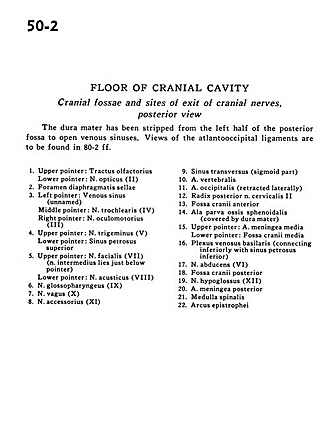
Floor of cranial cavity
Cranial fossae and sites of exit of cranial nerves, posterior view
The dura mater has been stripped from the left half of the posterior fossa to open venous sinuses. Views of the atlantooccipital ligaments are to be found in 80-2 ff.
- Upper pointer: Olfactory tract Lower pointer: Optic nerve (II)
- Sella turcica
- Left pointer: Venous sinus (unnamed) Middle pointer: Trochlear nerve (IV) Right pointer: Oculomotor nerve (III)
- Upper pointer: Trigeminal nerve (V) Lower pointer: Superior petrosal sinus
- Upper pointer: Facial nerve (VII) (intermediate nerve lies just below pointer) Lower pointer: Acoustic nerve (VllI)
- Glossopharyngeal nerve (IX)
- Vagus nerve (X)
- Accessory nerve (XI)
- Transverse sinus (sigmoid part)
- Vertebral artery
- Occipital artery (retracted laterally)
- Dorsal root cervical nerve II
- Anterior cranial fossa
- Lesser wing sphenoid bone (covered by dura mater)
- Upper pointer: Middle meningeal artery Lower pointer: Middle cranial fossa
- Basilar venous plexus (connecting inferiorly with inferior petrosal sinus)
- Abducens nerve (VI)
- Posterior cranial fossa
- Hypoglossal nerve (Xll)
- Posterior meningeal artery
- Spinal cord
- Arch of axis