
Bassett Collection of Stereoscopic Images of Human Anatomy
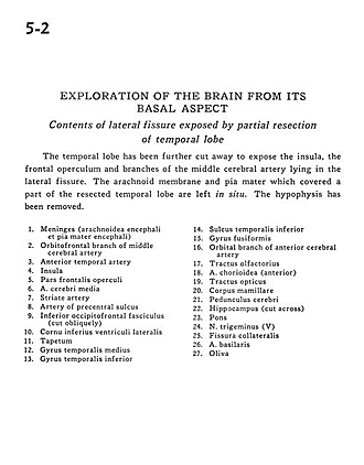
Exploration of the brain from its basal aspect
Contents of lateral fissure exposed by partial resection of temporal lobe
Image #5-2
KEYWORDS: Brain, Frontal lobe, Meninges, Telencephalon, Vasculature.
Creative Commons
Stanford holds the copyright to the David L. Bassett anatomical images and has assigned Creative Commons license Attribution-Share Alike 4.0 International to all of the images.

For additional information regarding use and permissions, please contact the Medical History Center.

Exploration of the brain from its basal aspect
Contents of lateral fissure exposed by partial resection of temporal lobe
The temporal lobe has been further cut away to expose the insula, the frontal operculum and branches of the middle cerebral artery lying in the lateral fissure. The arachnoid membrane and pia mater which covered a part of the resected temporal lobe are left in situ. The hypophysis has been removed.
- Meninges (arachnoid and pia mater encephali)
- Orbitofrontal branch of middle cerebral artery
- Anterior temporal artery
- Insula
- Frontal part of operculum
- Middle cerebral artery
- Striate artery
- Artery of precentral sulcus
- Inferior occipitofrontal fasciculus (cut obliquely)
- Inferior horn of lateral ventricle
- Tapetum
- Medial temporal gyrus
- Inferior temporal gyrus
- Inferior temporal sulcus
- Fusiform gyrus
- Orbital branch of anterior cerebral artery
- Olfactory tract
- Choroidal artery (anterior)
- Optic tract
- Mamillary body
- Cerebral peduncle
- Hippocampus (cut across)
- Pons
- Trigeminal nerve (V)
- Collateral fissure
- Basilar artery
- Olive