Bassett Collection of Stereoscopic Images of Human Anatomy
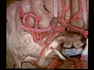
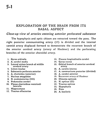
Exploration of the brain from its basal aspect
Close-up view of arteries entering anterior perforated substance
Image #5-1
KEYWORDS: Brain, Cerebellum, Medulla, Midbrain, Pons, Vasculature.
Creative Commons
Stanford holds the copyright to the David L. Bassett anatomical images and has assigned Creative Commons license Attribution-Share Alike 4.0 International to all of the images.

For additional information regarding use and permissions, please contact the Medical History Center.
Exploration of the brain from its basal aspect
Close-up view of arteries entering anterior perforated substance
The hypophysis and optic chiasm are retracted toward the pons. The right posterior communicating artery (17) is divided and the internal carotid artery displaced forward to demonstrate the recurrent branch of the anterior cerebral artery (artery of Heubner) and the perforating branches of the anterior choroidal artery.
- Orbital gyrus
- Middle cerebral artery
- Lateral striate branch of middle cerebral artery
- Entrance to insula
- Anterior perforated substance
- Choroidal artery (anterior)
- Amygdaloid nucleus
- Oculomotor nerve (III)
- Cerebral peduncle
- Choroid plexus lateral ventricle
- Hippocampus
- Olfactory tract
- Longitudinal fissure (cerebral)
- Straight gyrus
- Orbital branch of anterior cerebral artery
- Internal carotid artery
- Posterior communicating artery (divided)
- Anterior cerebral artery
- Recurrent artery of Heubner
- Optic chiasm
- Optic nerve (II)
- Optic tract
- Hypophysis
- Pons
- Basilar artery