Bassett Collection of Stereoscopic Images of Human Anatomy
General orientation views of dissection
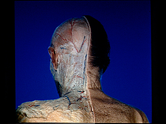
Superficial structures of head and neck, posterior view
Image #49-2
KEYWORDS: Connective tissue, Muscles and tendons, Peripheral nervous system, Vasculature, Fascia and connective tissue, Overview.
Creative Commons
Stanford holds the copyright to the David L. Bassett anatomical images and has assigned Creative Commons license Attribution-Share Alike 4.0 International to all of the images.

For additional information regarding use and permissions, please contact the Medical History Center.
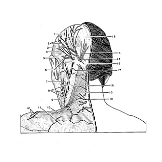
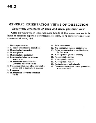
General orientation views of dissection
Superficial structures of head and neck, posterior view
Close-up views which illustrate more details of this dissection are to be found as follows
- Galea aponeurotica
- Occipital artery (lateral branches)
- Superior auricular muscle
- Occipitalis muscle
- Posterior auricular muscle
- Posterior auricular lymph nodes
- Sternocleidomastoid muscle (covered by supeficial fascia)
- Cutaneous filaments of lesser occipital nerve and greater auricular nerve
- Platysma
- Trapezius muscle (covered by superficial fascia)
- Superficial fascia
- Posterior supraclavicular nerves
- Galea aponeurotica virtually absent in this area
- Occipital artery (medial branch)
- Third occipital nerve
- Greater occipital nerve
- Lesser occipital nerve
- Posterior cervical triangle
- Cutaneous branch of posterior branch cervical nerve V