Bassett Collection of Stereoscopic Images of Human Anatomy
Osteology
Right upper molar and premolar teeth, lateromedial roentgenogram
Image #47-4
KEYWORDS: Bones cartilage joints, Face, Mouth.
Creative Commons
Stanford holds the copyright to the David L. Bassett anatomical images and has assigned Creative Commons license Attribution-Share Alike 4.0 International to all of the images.

For additional information regarding use and permissions, please contact the Medical History Center.
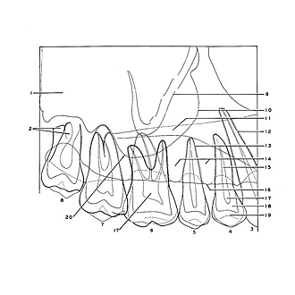
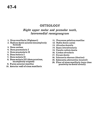
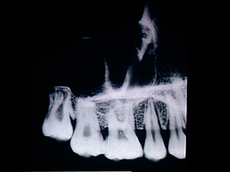
Osteology
Right upper molar and premolar teeth, lateromedial roentgenogram
- Maxillary sinus (Highmore)
- Roots molar III (serotini) (incompletely formed)
- Canine
- Premolar I
- Premolar II
- Molar I
- Molar II
- Molar III (serotinus incompletely erupted)
- Anterior surface of maxilla
- Anterior wall of maxillary sinus
- Palatine process of maxilla
- Root of canine
- Alveolus
- Interalveolar septa
- Root canal
- Alveolar border
- Cavity
- Substantia ebumea (dentine)
- Substantia adamantina (enamel)
- Floor of maxillary sinus (note close proximity to dental alveoli)