Bassett Collection of Stereoscopic Images of Human Anatomy
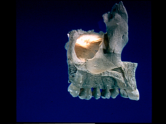
Osteology
Left maxilla, medial view
Image #45-2
KEYWORDS: Bones cartilage joints, Face, Mouth.
Creative Commons
Stanford holds the copyright to the David L. Bassett anatomical images and has assigned Creative Commons license Attribution-Share Alike 4.0 International to all of the images.

For additional information regarding use and permissions, please contact the Medical History Center.
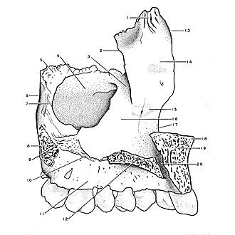
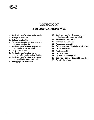
Osteology
Left maxilla, medial view
- Articular surface for frontal bone
- Lacrimal margin
- Lacrimal sulcus
- Maxillary sinus, visible through maxillary hiatus
- Articular surface for orbital process palatine bone
- Body of maxilla
- Articular surface for perpendicular part palatine bone
- Articular surface for pyramidal process palatine bone
- Pterygopalatine sulcus
- Articular surface for horizontal process palatine bone
- Alveolar process
- Palatine process
- Frontal process
- Crista ethmoidalis (faintly visible)
- Conchal crest
- Nasal surface
- Nasal incisure
- Anterior nasal spine
- Articular surface for right maxilla
- Incisive canal