
Bassett Collection of Stereoscopic Images of Human Anatomy
Creative Commons
Stanford holds the copyright to the David L. Bassett anatomical images and has assigned Creative Commons license Attribution-Share Alike 4.0 International to all of the images.

For additional information regarding use and permissions, please contact the Medical History Center.

Osteology
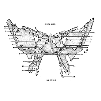
Sphenoid bone, anterior view
- Greater wing
- Lesser wing
- Frontal margin
- Orbital surface
- Zygomatic margin
- Aperture of sphenoidal sinus
- Sphenoidal crest
- Foramen rotundum
- Upper pointer: Sphenomaxillary surface Lower pointer: Rostrum of sphenoid
- Upper pointer: Medial plate of pterygoid process Lower pointer: Pterygopalatine sulcus
- Articular surface for pyramidal process of palatine bone
- Pterygoid fissure
- Groove (often a foramen) for recurrent meningeal branch of lacrimal artery
- Location of optic foramen
- Superior orbital fissure
- Temporal fossa
- Articular surface for ethmoid bone
- Sphenoidal concha
- Infratemporal fossa
- Pterygoid canal
- Processus vaginalis
- Angular spine
- Lateral plate of pterygoid process
- Pterygoid hamulus