
Bassett Collection of Stereoscopic Images of Human Anatomy
Creative Commons
Stanford holds the copyright to the David L. Bassett anatomical images and has assigned Creative Commons license Attribution-Share Alike 4.0 International to all of the images.

For additional information regarding use and permissions, please contact the Medical History Center.

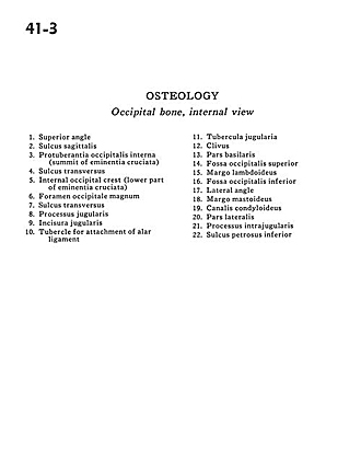
Osteology
Occipital bone, internal view
- Superior angle
- Sagittal sulcus
- Internal occipital protuberance (summit of cruciate eminence)
- Transverse sulcus
- Internal occipital crest (lower part of cruciate eminence)
- Foramen magnum
- Transverse sulcus
- Jugular process
- Jugular incisure
- Tubercle for attachment of alar ligament
- Jugular tubercle
- Clivus
- Basilar part
- Superior occipital fossa
- Lambdoidal margin
- Inferior occipital fossa
- Lateral angle
- Mastoid margin
- Condyloid canal
- Lateral part
- Intrajugular process
- Inferior petrous sulcus