
Bassett Collection of Stereoscopic Images of Human Anatomy
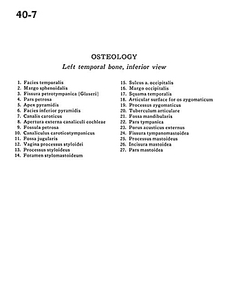
Osteology
Left temporal bone, inferior view
Image #40-7
KEYWORDS: Bones cartilage joints, Ear.
Creative Commons
Stanford holds the copyright to the David L. Bassett anatomical images and has assigned Creative Commons license Attribution-Share Alike 4.0 International to all of the images.

For additional information regarding use and permissions, please contact the Medical History Center.

Osteology
Left temporal bone, inferior view
- Temporal surface
- Sphenoidal margin
- Petrotympanic fissure
- Petrosal part
- Pyramidal apex
- Inferior pyramidal surface
- Carotid canal
- External opening cochlear canal
- Petrosal fossa
- Tympanic canaliculus
- Jugular fossa
- Sheath of styloid process
- Styloid process
- Stylomastoid foramen
- Sulcus occipital artery
- Occipital margin
- Temporal bone (squamous part)
- Articular surface for zygomatic bone
- Zygomatic process
- Articular tubercle
- Mandibular fossa
- Tympanic part
- External acoustic meatus
- Tympanomastoid fissure
- Mastoid process
- Mastoid incisure
- Mastoid part