Bassett Collection of Stereoscopic Images of Human Anatomy
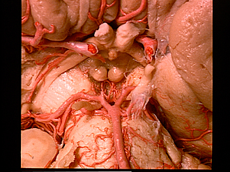
Exploration of the brain from its basal aspect
Arteries of interpeduncular fossa
Image #4-6
KEYWORDS: Bones cartilage joints, Brain, Cerebellum, Meninges, Peripheral nervous system, Vasculature, Ventricules.
Creative Commons
Stanford holds the copyright to the David L. Bassett anatomical images and has assigned Creative Commons license Attribution-Share Alike 4.0 International to all of the images.

For additional information regarding use and permissions, please contact the Medical History Center.
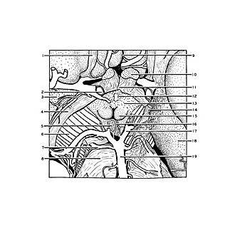
Exploration of the brain from its basal aspect
Arteries of interpeduncular fossa
The numerous branches of the posterior cerebral arteries are illustrated in this close-up view as they enter the posterior perforated substance in the depths of the interpeduncular fossa. A piece of arachnoid membrane covers the left third nerve and cerebral peduncle (right side of view). The space seen between this membrane and the brain represents a portion of the interpeduncular cistern.
- Olfactory tract right
- Internal carotid artery
- Anterior perforated substance
- Optic tract
- Interpeduncular fossa
- Cerebral peduncle
- Superior cerebellar artery
- Pons
- Longitudinal fissure (cerebral)
- Optic nerve (II)
- Optic chiasm
- Infundibulum
- Tuber cinereum
- Oculomotor nerve (III)
- Mamillary body
- Medial central branch of posterior cerebral artery
- Posterior cerebral artery
- Hippocampal gyrus
- Basilar artery